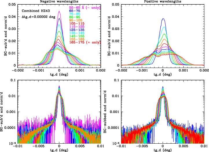
In order to cause the LSF structure seen in Fig. 12, errors in pixel positions would have to be at least 0.0002 deg, corresponding to errors of 5 or more pixels sustained over tens of Å (many cm on the HRC-S). Such large distortions would be readily apparent in work on the HRC-S degap map and in observed-vs-known source positions but are not seen.
The two outer plates are tilted by a few degrees to follow the Rowland circle (see POG Fig. 9.5--scroll up). Errors in those tilts would slightly degrade spectral resolution but not affect LSFs along tg_d. Rotations about the long axes of the plates would distort the LSFs but would have to be many degrees to have a noticeable effect and would not cause the observed LSF "bumpiness" seen in Fig. 12. Off-axis sources would also exhibit anomalously large PSFs, which are not seen. To be thorough, however, we analyzed data from ObsID 9617, an observation of Sirius B (a low energy continuum source like HZ43 but with a lower event rate) made with a Z offset pointing of about 400 pixels (~50"), placing the dispersed spectrum very near the boundary of the Low Energy Suppression Filter (LESF; see POG Fig. 7.1). If either HRC-S plate had a significant tilt about its long axis the off-axis LSFs of this observation would differ from those on axis.
The Sirius B observation dithered across the LESF boundary, losing approximately half of the X-ray events to absorption in the thicker Al coating of the LESF. To maximize S/N we therefore filtered out events with chipx>2600, leaving about 17 pixels less than half the 304-pixel dither pattern. Our comparison on-axis observation was ObsID 59 of HZ43. Using only a portion of the dither pattern should not significantly affect the LSFs but for consistency we used the same portion of the dither pattern in the HZ43 data processing. LSFs were then extracted in 10-Å bins as for Fig. 12, with proper subtraction of the sloping background caused by chipx filtering. As seen in Fig. C1, the profiles are essentially identical, confirming that any errors in HRC-S tilts are indeed negligible.
|
|
| Fig. C1. Comparison of tg_d profiles at long wavelengths from on-axis (HZ43) and Zoffset=50" (Sirius B) observations. |
Spectra dispersed by a single LETG facet are symmetric between + and - orders. Alignment of 3 facets that make up one grating module (see POG Figs. 9.1, 9.2, and 9.3) was usually better than ±15", corresponding to tg_d=±0.00007 deg around 160 Å. Alignment of the 180 modules that make up the LETG is accurate to TBD. An alignment error for a given facet will tilt the spectrum slightly but it will still be symmetric about 0th order for that facet. The overall symmetry of LETG spectra will be broken, however, if facets with different tilts do not have the same 0th order. This is shown schematically in Fig. C2. In fact, the Chandra mirrors, or HRMA shells, are known to focus in slightly different positions with additional variation between different portions of a given shell. The observed LSFs can therefore be explained by assuming there are several clusters of slightly different LETG facet alignments combined with a nonuniform distribution of those clusters across the HRMA shells.

|
| Fig. C2. Schematic of dispersed spectra with four groups of grating-facet tilts and 0th order positions (dots). The vertical axis (tg_d) is highly stretched. The spectrum from each group of facets is symmetric about its 0th order but the composite LETG spectrum will have asymmetry in both tg_d and wavelength. This figure illustrates the concept and does not represent the true LETG in detail, although the colored dots do mark the locations of the foci of the four HRMA shells at low energies: 1=red, 3=orange, 4=green, 6=blue, composite focus in black. Foci of shells 3 and 6 are separated in the tg_d direction by 5 μm, a little less than one pixel. There is also some spread in focus locations of different portions of a single shell so the total spread in Z is larger than 5 μm. |
Fig. C3 shows a rough attempt to match the observed LSFs at 8 wavelengths using 6 facet groupings, corresponding to 7 peaks in the LSFs. Each peak was modeled as a gaussian that widens as f(|wavelength|). The only free parameters in this model are the peak amplitudes, 0th order positions (spanning a range of 12 μm in Z, the tg_d direction) and tilts, ranging over ~2'. As can be seen, this crude attempt does not match very well but it should be apparent that the general concept is valid. (LSFs in fact become highly non-Gaussian at long wavelengths, but doing a proper fit with more accurate models is beyond the scope of this work.)

|
| Fig. C3. Rough modeling of observed LSFs assuming 7 groups of LETG facets with varying number of facets, tilts, and 0th order Z positions. |